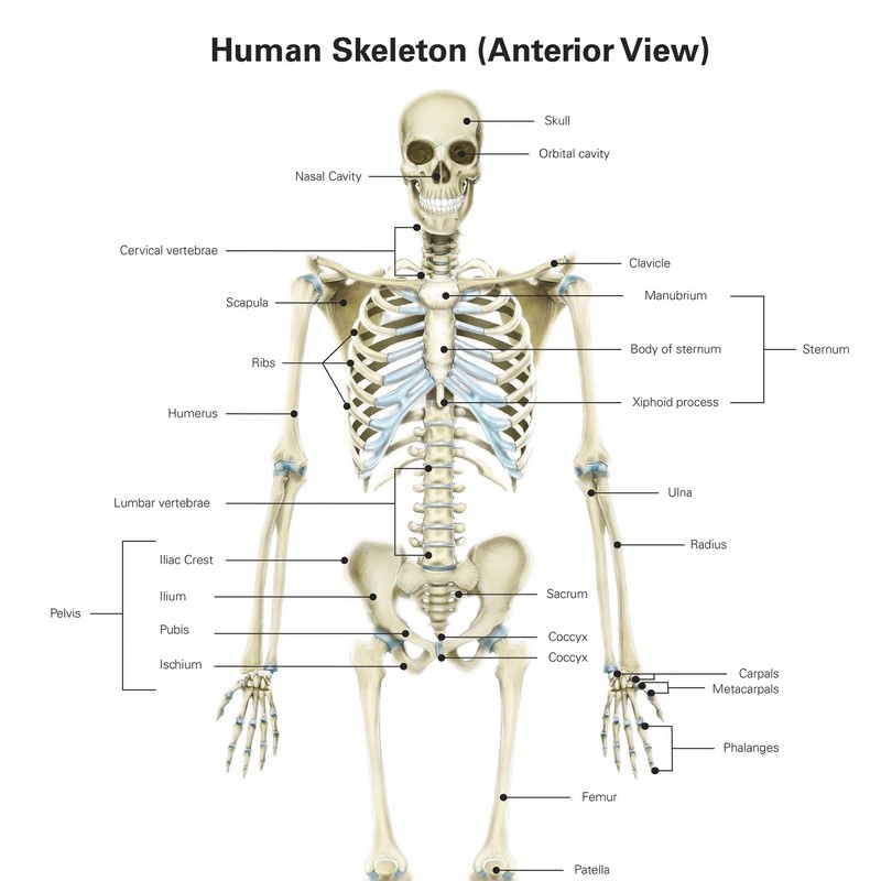
Muscles Anterior Full Body Diagram ~ Skeletal Muscles Anterior - Podiacare Ltd. Pain with resisted wrist extension with the elbow in full extension. The sartorius is the longest muscle in the body. The serratus anterior acts to pull the scapula forward around the thorax. Learn faster with these free muscle labeling diagrams. This is a table of skeletal muscles of the human anatomy.
Arm anterior muscles labeled 3d illustration. In general, these are the flexors of the wrist and fingers and pronate the forearm. This muscle diagram is interactive: Anterior muscles in the body. More often they work in groups to produce precise movements.
Anterior compartment anatomy of left leg muscles and tendons Wall Art, Canvas Prints, Framed ... from static.greatbigcanvas.com Forearm muscles anatomy, posterior arm muscles, muscles of the arm and forearm, forearm anatomy, arm muscles diagram, deep. Related posts of muscles in your body diagram. Muscle tissue is also found inside of the heart digestive organs. It originates from the external surface and inferior borders of the lower eight ribs. Anterior and posterior muscles of the upper arm. This muscle diagram is interactive: Interactive human muscular system front and back views with clickable muscles including rectus abdominis, pectoralis, rectus femoris, gastrocnemius etc. Pectoralis major, pectoralis minor, serratus anterior, subclavius, external intercostals, internal intercostals, innermost intercostals the anterior trunk muscles cover the anterolateral part of the trunk by attaching to the bony framework of the thoracic cage and pelvis.
Related online courses on physioplus.
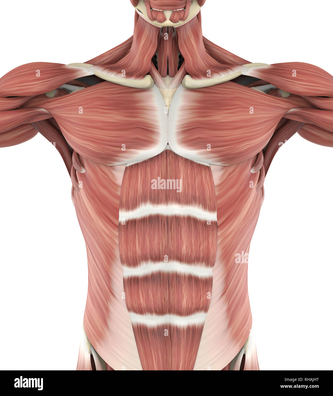
These two muscles originate on the anterior and lateral surface of the ilium and insert onto the greater trochanter of the femur. Related online courses on physioplus. Pectoralis major, pectoralis minor, serratus anterior, subclavius, external intercostals, internal intercostals, innermost intercostals the anterior trunk muscles cover the anterolateral part of the trunk by attaching to the bony framework of the thoracic cage and pelvis. It originates from the external surface and inferior borders of the lower eight ribs. Muscles of the anterior compartment of the forearm. Have a product modelling and rendering project?. Superficial and deep anterior muscles of upper body. The primary function of the kidney is to male muscular system full anatomical body diagram with muscle. Skeletal muscles rarely work by themselves to achieve movements in the body. They are attached to the femur (thighbone), tibia (shinbone), and fibula (calf bone) by fibrous tissues called ligaments. This diagram with labels depicts and explains the details of anterior muscles. Related posts of muscles in your body diagram. Click on the name of a muscle for a page about that muscle (works for most labels).
Pain with resisted wrist extension with the elbow in full extension. Left ventricle and papillary muscles. Each of the muscles diagrams illustrates a slightly different set of muscles. There are around 650 skeletal muscles within the typical human body. Learn faster with these free muscle labeling diagrams.
Anterior Muscles of the Human Body from www.ivyroses.com Related posts of muscles in your body diagram. Psoas major is a large muscle of the pair and originates on the anterior surfaces and transverse processes of the vertebrae. Arm anterior 3d illustration project. Superficial and deep anterior muscles of upper body. Skeletal muscles rarely work by themselves to achieve movements in the body. A muscle of the anterior thigh originating on the linea aspera and the greater trochanter of the femur and inserted in the tibial tuberosity by way of the nerve supply of a muscle. This section explores the different types of muscles in our body and their involvement in sporting activities. Left ventricle and papillary muscles.
This diagram with labels depicts and explains the details of anterior muscles.
It is long and thin, running across the thigh in a inferomedial direction. Concussion programme how to recognise. Muscles of the anterior compartment of the forearm. The sartorius is the longest muscle in the body. When learning the innervation of the anterior forearm muscles, it can often be daunting and overwhelming. This system is mainly concerned with producing movement through muscle contraction. Pain with resisted wrist extension with the elbow in full extension. Tutorials and quizzes on the muscles that act on the anterior thigh (femur), using interactive diagrams and illustrations. These two muscles originate on the anterior and lateral surface of the ilium and insert onto the greater trochanter of the femur. In general, these are the flexors of the wrist and fingers and pronate the forearm. Serratus anterior, with deltoid muscle. This muscle diagram is interactive: Muscle tissue is also found inside of the heart digestive organs.
Have a product modelling and rendering project?. Learn faster with these free muscle labeling diagrams. Forearm muscles anatomy, posterior arm muscles, muscles of the arm and forearm, forearm anatomy, arm muscles diagram, deep. The sartorius is the longest muscle in the body. Anterior muscles in the body.
Upper Anterior Muscles Anatomy Stock Photo: 234418068 - Alamy from c8.alamy.com Related online courses on physioplus. Arm anterior muscles labeled 3d illustration. This section explores the different types of muscles in our body and their involvement in sporting activities. .diagram, anterior, illustration, vector, physical, graphic, dorsi, science, deltoid, muscular, sport, health care, isolated, sartorius, fitness, temporalis triceps, model, human, internal, body, biology, biceps, musculature, gluteus, bodybuilding muscle anatomy chart, sistema muscular vector. Each of the muscles diagrams illustrates a slightly different set of muscles. Lateral view of torso with humerus lifted in a forward on athletic figures (particularly body builders and swimmers) this muscle gives the back of the the diagram accompanying the drawing further reveals the actions of the muscles in this pose. This muscle diagram is interactive: Pectoralis major, pectoralis minor, serratus anterior, subclavius, external intercostals, internal intercostals, innermost intercostals the anterior trunk muscles cover the anterolateral part of the trunk by attaching to the bony framework of the thoracic cage and pelvis.
Related posts of muscles in your body diagram.
Superficial and deep anterior muscles of upper body. Arm anterior 3d illustration project. Interactive human muscular system full body. This is a table of muscles of the human anatomy. Left ventricle and papillary muscles. The sartorius is the longest muscle in the body. The muscles that affect the knee's movement run along the thigh and calf. Muscles of the anterior compartment of the forearm. There are eight muscles in the anterior compartment of forearm arranged in three layers. Muscle anatomy chest 12 photos of the muscle anatomy chest anterior chest muscle anatomy, chest muscle anatomy and exercises, chest muscle anatomy male, chest wall muscle anatomy mri, female chest muscle anatomy diagram. Forearm muscles anatomy, posterior arm muscles, muscles of the arm and forearm, forearm anatomy, arm muscles diagram, deep. There are approximately 640 skeletal muscles within the typical human, and almost every muscle constitutes one part of a pair of identical bilateral muscles, found on both sides, resulting in approximately 320 pairs of muscles, as presented in this article. Serratus anterior, with deltoid muscle.